Learning neuroanatomy terms is a lot like learning a second language.
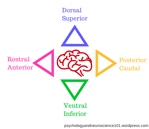
In neuroanatomy, we don’t say top, bottom, back and front. Instead, we have a whole different range of terminology for describing which region of the brain we are talking about.
But don’t worry, with this blog post, learning all the new names will be a doddle!
Top
The “top” of the brain is referred to as either the:
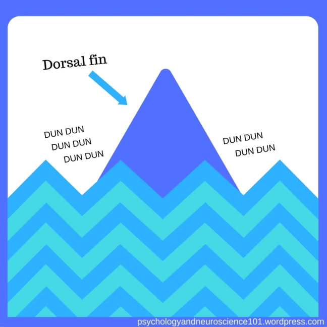
Dorsal
Superior
…parts of the brain.
Remember: Dorsal
To remember that the top section of the brain is sometimes referred to as the dorsal region of the brain, think of the dorsalfin of a shark.
The dorsal fin sits at the top of the shark’s body.
This is the fin that often pokes out of the water before a shark attack in the movies.
Remember: Superior
To remember that the top section of the brain is sometimes referred to as the superior region of the brain, picture someone who thinks that they are superior to those around them, such as a mean queen.
You could say that they believe that they are above everyone else – just like the superior region of the brain is above all the other regions in the brain.
Image: from clipartwolf.com
Bottom
The bottom of the brain is referred to as either the:
Ventral
Inferior
…parts of the brain.
Remember: Ventral
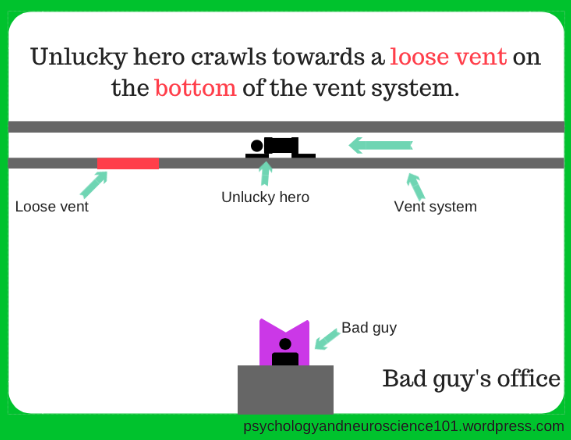
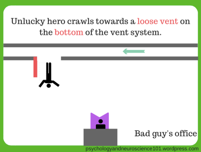
Ventral comes from the Latin “venter” meaning, belly.
To remember that the bottom section of the brain is sometimes referred to as the ventral region, think of how, in the movies, whenever the hero is moving through a ventilation system, they’ll usually end up stepping on a loose vent panel and falling right into the bad guy’s main office below.
Remember: Inferior
To remember that the bottom section of the brain is sometimes referred to as the inferior region, picture someone who thinks that they are inferior to those around them, such as sulky Sam.
Image: from clipartpanda.com
Sulky Sam is upset because Suzie got an A on her psychology test, whereas, he got a C.
Image: image of girl and boy both adapted from clipartpanda.com
Sulky Sam thinks he is inferior to Suzie.
Back
The back of the brain is referred to as either the:
Posterior
Caudal
…parts of the brain.
Remember: Posterior
To remember that the back of the brain is referred to as the posterior region, remember that the word posterior can also be another name for your butt. Therefore, you can think of the posterior region of the brain, as the brain’s butt or backside.
Remember: Caudal
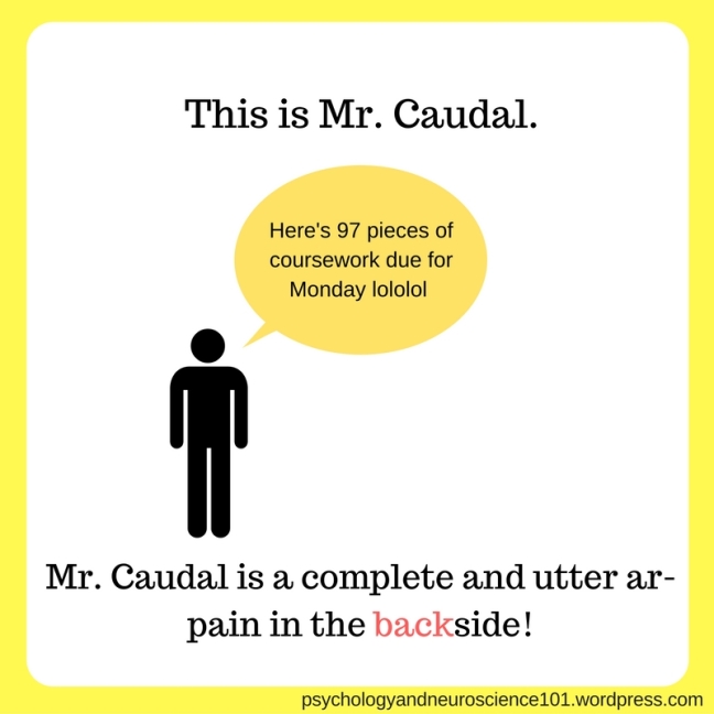
To remember that the back region of the brain is also sometimes referred to as the caudal region of the brain, imagine a really mean lecturer named Mr. Caudal who always sets loads of assignments.
You could say that Mr. Caudal is a pain in the backside!
(Remember- the ruder something is, the more likely you are to remember it!)
DISCLAIMER: Characters featured in this blog are not based on any persons, whether living, deceased, fictional or otherwise.
Front
The front of the brain is referred to as either the:
Rostral
Anterior
…parts of the brain.
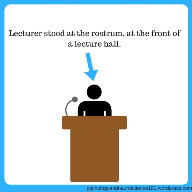
Remember: Rostral
To remember that the front section of the brain is sometimes referred to as the rostral region, remember that in a lecture, the speaker stands at a podium at the front of the lecture hall – this podium is called the rostrum.
The podium that a conductor uses when he stands in front of an orchestra is also called a rostrum.
Remember: Anterior
Image source: @antanddec at Twitter.com
To remember that anterior region of the brain is at the front of the brain, think of the famous television duo, Ant and Dec.
When you say Ant and Dec, Ant’s name always comes in front of Dec’s. You never say Dec and Ant, that would just be weird.
So, Ant is always in front of Dec – he is ANTerior to him.
In this post, we will look at how two neurons send a message between each other.
Just like two people can be said to share a chemical attraction, the process of two neurons sharing information is also a chemical affair.
Synapse
The synapse occurs at the point where the terminal button of one neuron meets the membrane (dendrite or soma) of another neuron.
The is a small gap between the synapse and the membrane known as the synaptic cleft. Information travels through A’s synapse, across the synaptic cleft and into the receptors on neuron B.
Synaptic transmission
Synaptic transmission basically refers to the delivery of a message via a synapse.
Synaptic transmission is a chemical process (as mentioned above) that deals with neurotransmitters.
Here is the process of what happens during synaptic transmission:
Binding
Let’s take a closer look at exactly what happens at step 7, when the neurotransmitter binds to the post-synaptic cell. (The post-synaptic cell is basically the one that’s doing the receiving).
The receptors in the membrane of the post-synaptic cell contain these things called ion channels. You can picture these as doors into/out of the cell.
These ion channels either let ions in or out of the postsynaptic cell.
The method in which these ion channel doors are opened involves things called neurotransmitters. The role of a neurotransmitter is to bind themselves to a receptor’s binding site. This will let the receptor’s ion channel will open and allow ions to move either in or out of the postsynaptic cell.
So, think of the neurotransmitters as being little keys and the binding sites on the receptors are the locks. Binding to a binding site unlocks the door and opens the ion channel door.
However, there’s a catch, each neurotransmitter will only bind to one certain type of receptor. For instance, one neurotransmitter may only bind to a binding site on a receptor that handles Na+ ions (i.e. one with an ion channel that lets Na+ ions into the cell). Another may only bind to a binding site on a receptor that handles K+ ions, etc.
This is called a lock and key system. Each neurotransmitter only binds to one type of receptor, in the same way that one key can only unlock one lock.
Remember, letting ions into and out of the cell creates a certain postsynaptic potential (fancy name for the receiving cell’s charge).
The postsynaptic potential that is created (excitatory or inhibitory) depends on which ion channel (door) is unlocked and thus, which ions are moving into/out of the cell.
You can have two types of postsynaptic potential:
EPSP = excitatory postsynaptic potential
IPSP = inhibitory postsynaptic potential
An EPSP basically means that the receiving neuron cell has been encouraged to fire and send on the message to other neurons.
So, if your neurons were passing along this message: “JUMP”, a specific ion channel would be opened on the postsynaptic cell that would allow a certain type of ion through that would result in the creation of an EPSP.
This would encourage the neuron to fire and pass on the message to other neurons, leading to the behaviour eventually being carried out.
Whereas, an IPSP means that a receiving neuron has been discouraged from firing and sending on the message.
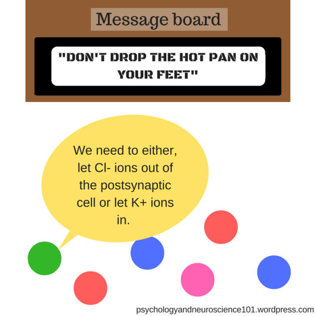
So, if the message were “DON’T OPEN YOUR HAND AND DROP THE BOILING PAN OF WATER”, then a different ion channel would open on the postsynaptic cell that would allow a different type of ion to enter/leave the cell, thus creating an IPSP.
This would prevent the neuron from firing and thus, stop you from opening your hand and dropping the pan.
But which ions create an EPSP and which create an IPSP?
I’m glad you asked.
Na+
Na+ can only move INTO the postsynaptic cell and not out. If a whole lot of Na+ does enter the postsynaptic cell then an EPSP is created. This means that the cell will become depolarised (when the postsynaptic cell’s charge moves closer to zero).
Try imagining that the postsynaptic cell is in financial debt and the Na+ ions are a form of currency. As more Na+ ions enter the cell, the cell’s outlook on life becomes less negative.
Also, you can remember that depolarised = moves closer to zero because polarised = gains electric charge, so depolarised must mean that it is losing that charge.
K+
K+ can only move OUT of the postsynaptic cell. If a lot of K+ does exit the cell, then an IPSP is caused in the postsynaptic cell. This hyperpolarises the postsynaptic cell, making it more negative.
Imagine that the post-synaptic cell is upset that all the K+ ions are leaving, like it’s losing it’s babies, so as a result it has a more negative outlook on life.
Cl-
Cl- can only move INTO the postsynaptic cell. If lots of Cl- enters the postsynaptic cell, then an IPSP is caused in the postsynaptic cell. This hyperpolarises the postsynaptic cell, so that it becomes more negative.
Imagine that the postsynaptic cell is enjoying its life, minding it’s own business, then a hoard of Cl- ions move into it’s membrane, disturbing its peace. This leaves the postsynaptic cell feeling rather negative. Like when your younger siblings burst into your room and ruin your peaceful time.
Ca2+
This one is a sneaky one. He’s very different from the rest. Be sure to look out for him. Basically, Ca2+ can only move INTO the cell and once it is in there it’s main job is to activate enzymes. Forget all that IPSP and EPSP nonsense, Ca2+ ain’t go time for that.
In summary:
Confused?
Let’s go through it again from the top:
(NT = neurotransmitter)
So, as you can see, the neurotransmitters work as keys to unlock the ion channel doors on the receptors, so that ions can pass in and out of the cell. As ions pass in and out of the cell, the appropriate PSP (postsynaptic potential) is created that either encourages or discourages a given behaviour from happening.
Receptors
Receptors sit on the edge of the post-synaptic cell and their job is to receive the incoming ions/messages.
Receptors come in two types:
ionotropic receptors
metabotropic receptors
Both types have binding sites. Binding sites are the points where the neurotransmitter attaches to the receptor, thus opening the ion channel and allowing ions to enter the post-synaptic cell.
Ionotropic receptor (eye on oh trow pik) – Direct method
With an ionotropic receptor, the ion channel on the receptor will only open when a neurotransmitter binds to that receptor’s binding site.
Think of the neurotransmitter as a key to open the receptor’s “ion channel” door. The binding site would then be the lock.
This method is good for things that you need to be updated quickly, like your sense of sight and hearing.
The use of metabotropic receptors makes up the indirect method of transmission between neurons.
This is how metabotropic receptors work:
Here’s a plain text version:
NT binds to the ion channel (NT= neurotransmitter)
This activates the G-protein
G-protein activates an enzyme
Enzyme produces second messengers (blue arrows on diagram)
Second messengers open the ion channels
So, as you can see, they work using a chain reaction.
This chain reaction means that postsynaptic potentials (PSPs) are produced slower than those made by ionotropic receptors.
This is good for stuff that you need to last for a while, for instance, sense of taste, smell and pain.
You can try and remember that metabotropic receptors form the indirect method of transmission between neurons by associating “metabotropic” with the term “meta”.
When a piece of creative work is termed as meta, it usually means that it indirectly refers to itself or it’s genre/industry. An example of a meta-movie could be a movie about people making their own movie about the film industry. Therefore, the meta-movie would be an indirect comment on itself/it’s industry.
Thus, we can think metabotropic = meta = indirect method.
Likewise, we can then remember that ionotropic receptors must be the direct method by default.
Termination
Let’s look more closely at how the excess neurotransmitter left outside the cell is dissipated away.
This dissipation mainly happens via two ways:
1. Enzymatic deactivation/degradation
The neurotransmitter is eventually broken down by an enzyme into other substances.
e.g., acetylcholinesterase (a see tul koh lin ess ter ace) breaks down Ach into choline and acetic acid.
2. Reuptake
The excess neurotransmitter is taken back into the presynaptic terminal (the cell it just came out of).
Neural integration
The excitatory PSPs (postsynaptic potentials) increase the chance that the neuron will fire.
Whereas, the inhibitory PSPsdecrease the chance that the neuron will fire.
Integration is basically the fancy term for when you take the excitatory PSPs then take away the inhibitory PSPs.The total postsynaptic potential from that equation determines whether the neuron will actually fire or not.
(Note: the numbers in the picture above are meaningless and are merely to illustrate the integration process)
Important points:
It is important to remember that inhibitory PSPs don’t always inhibit behaviour.
If there’s an inhibition of inhibitory neurons then there’s a greater chance of behaviour occurring .
E.g. So, an inhibitory neuron may be stopping you dropping a heavy bowling ball that you’re carrying onto your friend’s foot. Use an IPSP to inhibit those neurons (turn them off) and you’ll have one less friend…and your friend will have one broken foot.
If there’s an excitation of inhibitory neurons then there’s less chance of behaviour occurring.
E.g. So, another inhibitory neuron might be stopping you from dropping a pan of boiling water onto your foot. Luckily for you, using an EPSP to excite this neuron boosts its inhibiting power meaning that you are extremely unlikely to drop the pan.
Think of EPSPs and IPSPs like potions or pills. If an inhibitory neuron takes an EPSP pill, it becomes excited and does its job of inhibiting behaviour twice as hard. If an inhibitory neuron takes an IPSP pill, it becomes inhibited and is less effective at its job of inhibiting behaviour.
Neurotransmitters
The following neurotransmitters are all actually amino acids.
Let’s look at some of the main neurotransmitters and what they do:
Glutamate
Glutamate is the most common excitatory neurotransmitter floating around in your CNS (central nervous system).
It’s unique in that it has the ability to bind to a large number of receptors.
Glutamate is particularly important for learning and memory processes.
You can remember this because you can say that glutamate makes information stick in your head like glue.
GABA (gamma-aminobutyric acid)
GABA is the most common inhibitory neurotransmitter in your CNS.
This means that GABA’s main job is to lower the likelihood of a neuron firing.
Basically, GABA is a massive party-pooper.
Acetylcholine (ACh) (a see tul koh leen)
Image credit: muscle image from Clker.com
ACh is a neurotransmitter that is located in both the CNS and the PNS (peripheral nervous system).
In particular, ACh is usually located at neuromuscular junctions.
Hence, ACh has a big role in muscle contraction.
Some poisons have been created that exploit the use of ACh in the CNS:
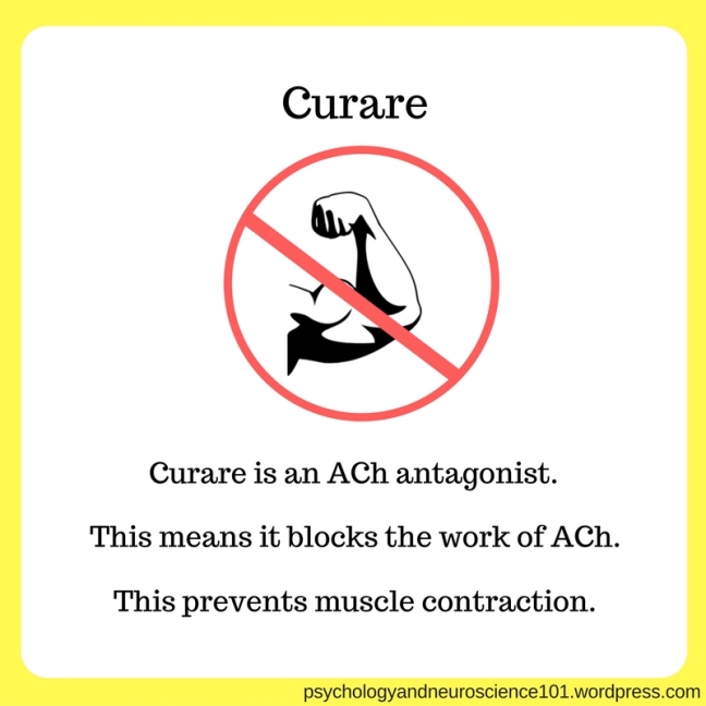
Curare (kew rahr ee)
Image credit: muscle image from Clker.com
Curare is an ACh antagonist. This means that it blocks the functioning of ACh.
By doing this, curare effectively prevents muscle contraction from happening, leaving the victim paralysed.
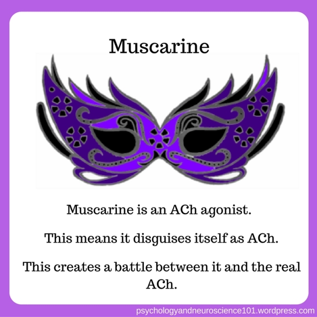
Muscarine
Image credit: mask from Clker.com
Muscarine is an ACh agonist. This means that is disguises itself as ACh, thus creating a battle between itself and the genuine ACh at binding sites.
Uses for these poisons
These two poisons have actually been put to use in:
hunting (by using darts coated in curare to paralyse a target)
surgery (as a local anaesthetic)
Monoamines
The following neurotransmitters are known as the monoamines.
They are made by the neurons in your brain.
They are also G-protein coupled (so, they have a G-protein attached to them. Don’t worry too much about this – just remember it).
The monoamines can be either excitatory or inhibitory, it all depends on which receptor subtype they bind to. In a sense, they’re kind of like chameleons.
Let’s take a look at two member of the monoamine family and what jobs they do:
Dopamine:
Think of dopamine as a drug baron.
The things it cares about are:
motor control (being in control of their crew, or your body in dopamine’s case)
reward (such as cold, hard cash)
addiction (dopamine explains why many drugs are so addictive)
Looking at it like that, it would be easy to picture a dopamine as a greedy, cruel drug baron. We could even call him Donny “the drug lord” Dopamine.
Image credit: Clker.com
Serotonin:
Serotonin is another member of the monoamine family.
Serotonin is basically a stereotypical mum. It has many jobs to do in order to keep you functioning, these include:
Checking that you’re feeling okay (mood regulation)
Making sure that you’re eating right (regulating eating)
Making sure that you’re getting enough sleep (regulating sleep)
Making sure that you’re paying attention when crossing the street, etc (regulating arousal)
Letting you know when to stop doing something before you get really badly hurt (experience of pain)
This post will focus on the transmission of information within a single neuron, i.e. how information moves through a single neuron.
Transmission within a neuron is an electrical process (which is the focus of this post).
Transmission between two different neurons is a chemical process (the focus of this other post that you can check out here).
How to remember this is seen here:
So, the attraction between Geoff and Suzie is chemical. Much like how the transmission between two different neurons is also chemical (chemical process).
The surge that Geoff felt within his own heart was electric. Much like how the transmission within a single neuron is also electric (electrical process).
Every cell in your body will carry an electrical charge
However, whether this charge is positive or negative differs between the inside and the outside of the cell.
All cells can be thought of as Regina George from the movie “Mean Girls”. If you haven’t seen Mean Girls then think of a person who smiles at your face but secretly hates you.
Like Regina George, cells are positive on the outside but negative on the inside.
Resting potential
First, let’s look at what actually makes up a cell:
The membrane carries an electrical charge known as the membrane potential.
The membrane potential is essentially the difference between the electrical potential inside the cell and the electrical potential outside the cell:
The membrane potential of the cell when at rest (when everyone is just chilling and no info is being transmitted) can also be called the resting potential.
For a neuron, the resting potential is -70Mv.
Neurons send information to other neurons by temporarily altering the their overall polarity (electrical charge).
Membrane potential is altered by the movement of ions in and out of the cell.
The ions are prevented from moving in and out of the cell by these two forces:
Diffusion
Electrostatic pressure
Here’s a reminder of what those two forces actually are:
Diffusion: molecules (water in the example) move from an area of high concentration to an area of low concentration.
Electrostatic pressure: the electric charge of each molecule determines whether two molecules will attract or repel each other.
Ions
What are ions anyway?
The cell contains many charged molecules called ions.
Ions come in two flavours:
Cation (+vely charged ions)
Anion (-vely charged ions)
The fluid inside the cell is called the intercellular fluid. This contains potassium (K+) ions.
The fluid outside the cell is called the extracellular fluid. This contains sodium (Na+) and chloride (Cl-) ions.
The ions at resting potential
“What’s this about K+, Cl- and Na+?”, I hear you ask.
To understand more about these ions and what role they play in the transmission of information within a single neuron, let’s look at how they behave at resting potential.
At resting potential, no information is being sent down the neuron, so the ions do not move. You can think of this as their default state.
To understand how they all behave at rest, we are going to imagine that this cell is actually a nightclub, because what else do ions do when they aren’t working but go clubbing?
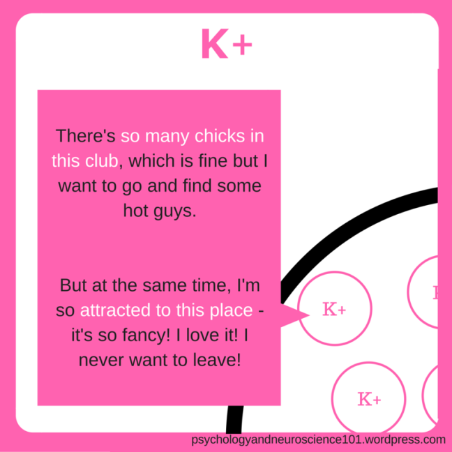
K+ (inside)
Diffusion: means that, although the K+ ions aren’t moving yet, they WANT to move out of the cell, which has a lower concentration of K+ ions than the inside of the cell.
BUT
Electrostatic pressure: means that the K+ ions are attracted to negative inside of the cell.
Cl- (outside)
Diffusion: means that the Cl- ions want to move in to the cell, which has a lower concentration of Cl- ions than the outside of the cell.
BUT
Electrostatic: means they are repelled by the negative inside of the cell.
Na+ (outside)
Diffusion: means that the Na+ ions want to move in to the cell, which has a lower concentration of molecules than the outside of the cell.
AND
Electrostatic: means that they are attracted to the negative inside of the cell.
So, all in all:
Sodium-potassium pumps
Okay, so eventually, a lot of Na+ ions travel into the cell and quite a few lucky K+ ions manage to sneak out as the concentration inside the cell increases.
Now, what happens next?
Well, that’s where the sodium-potassium pumps come in. Their purpose is to pump all the Na+ ions out of the cell and pump the escaped K+ back into the cell.
To remember this, let’s go back to the nightclub analogy:
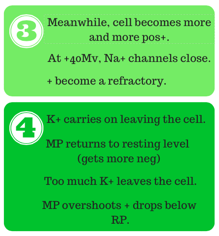
So, that’s how the ions behave at rest. However, when a piece of information needs to be sent down a neuron, the neuron undergoes a rapid change in electrical charge. This new charge is called the action potential.
It’s like on weekends when it’s just chilling, the neuron has a resting potential. But when it need to work and send signals, it has an action potential.
Action potential
An action potential is a rapid change in the polarisation (electrical charge) of the neuron in order to send a signal down said neuron.
For this signal to be sent, the neuron must first gain enough action potential to reach the threshold of excitation.
Think of the threshold of excitation as a big red button. The neuron must have reached a certain action potential in order to push this big red button.
Pushing the big red button, i.e. reaching the threshold of excitation, then causes the neuron to fire it’s signal down the axon by allowing the ions to finally move in and out of the cell.
This is an “all or nothing” process, the neuron either fires or it doesn’t.
During the process of the neuron firing, voltage-dependent ion channels open or close depending on the current membrane potential of the neuron (hence, voltage-dependent). These ion channels are what allow the ions to enter/leave the neuron, they’re like little doors really.
At various parts of the firing process, the neuron is depolarised and hyperpolarised:
Depolarisation: decrease from normal resting potential (overall charge moves closer to zero) – if polarisation means electrical charge then de-polarisation means less charged.
Hyperpolarisation: increase in action potential (the overall charge becomes more negative).
Here’s the process of the neuron firing:
Now let’s look at the process in more detail:
The point of becoming a refractory is to stop the neuron from firing a second time since it is still over the threshold of excitation (see above graph).
Saltatory conduction
The gaps in myelin sheath are called the nodes of Ranvier.
As the action potential travels down the axon, it is regenerated at these gaps. This makes it look like the action potential is jumping down the axon.
This process is called saltatory conduction because the word saltare means to “to jump“.
But why?
This process allows for:
Fast conduction – travels down axon faster.
More energy efficient
Under the myelin sheath, decremental conduction occurs. This is when the action potential decreases as it moves down the axon. However, the action potential is then regenerated to its full strength at each node of Ranvier. Therefore, saltatory conduction is energy efficient.
So, it looks like the full strength/size action potential is jumping down the axon:
First things first, let’s start with what a neuron actually is.
GCSE Biology coming at you in 3…2…1…
Image source: edited from pixabay.com
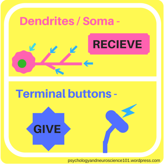
Soma – this is the part of the cell that contains the nucleus
Dendrites – these receive messages from other neurons
Axon – carries the message from the soma to the terminal buttons
Terminal buttons – sends the message on to the next neuron. These sent info to another neuron’s dendrites or soma.
Myelin sheath – protects the axon / helps send the message down the axon (more on that later).
You can remember that dendrites RECEIVE messages and terminal buttons SEND messages by thinking of it like this:
Dendrites look like the roots of a tree. A tree’s roots suck up all the water and nutrients from the soil. Therefore, the dendrites suck up all the information from the other neuron.
Whereas, the terminal buttons look a bit like the end of a creepy alien finger. So, you could imagine the process of sending a message from one neuron to another as a creepy game of tag, with the terminal buttons as the fingers that are about to tag another neuron.
Different types of neuron
Neurons actually come in 3 flavours:
The one you’re probably most familiar with is called the multipolar neuron.
Multipolar neurons are identifiable by the fact that they have:
one axon
many dendrites
This is easy to remember because multi- means multiple and this neuron has multiple dendrites.
Next we have the bipolar neuron.
A bipolar neuron has:
one axon
one dendrite tree
To remember this just imagine that the multipolar neurons are pansexual and like multiple genders (or rather they have multiple ends – because multiple dendric trees).
Whereas, a bipolar neuron can be thought of like a bisexual – they like 2 genders only (or rather they have 2 ends – one being a dendric tree, one being not)
Bipolar neurons are usually located in the visual/auditory systems (sensory systems).
The final neuron is a unipolar neuron.
A unipolar neuron has:
one axon – divided by two branches
one branch receives sensory information
the other branch sends information to the CNS
You can think of these as heterosexual neurons. All the information goes straight (sorry) through them. One branch receives info and the other branch sends it on to the CNS.
Connections
image source: pixabay.com
Remember that the terminal buttons are like alien fingers that send the information. Like when, after rubbing your feet on the ground, you touch someone to give them an electrostatic shock.
Also, remember that the dendrites are like tree roots that suck up the information from the terminal buttons.
Terminal buttons can also attach on to the soma, which can also receive information in the same way that the dendrites can.
Afferent and efferent neurons
The term “structure”refers to any part of the brain, e.g. the thalamus.
An afferent neuron is one that carries information to the structure.
Whereas, an efferent neuron is one that carries information away from the structure.
So, with an Afferent neuron – info Arrives at the structure.
And with an Efferent neuron – info Exits the structure.
Supporting cells
A great supporting cast is essential is you’re an actor…or a neuron.
This supporting cast goes by the collective name of the Glia (“glee ah”).
The glial cells include:
astrocytes (‘astro-sites’)
oligodendrocytes (‘oh li go den droh sites‘)
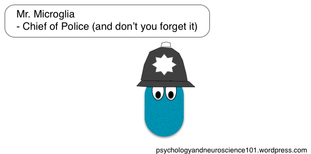
microglia (‘micro-glee-ah‘)
General function:
support the neurons
take away waste
give nutrients to the neurons
Astrocytes
Astrocytes are star-shaped. You can remember that because words beginning with astro- usually relate to space/stars (e.g. astronaut, astrology, astrophysics).
Astrocytes have two jobs:
removal of waste
synchronising neurons
Think of it like this:
Every astrocyte works two jobs. By day, Mr. Astrocyte is everybody’s favourite neighbourhood bin/garbage-man.
But in the evenings, Mr. Astrocyte coaches a team of synchronised swimmers and gets very competitive.
Oligodendrocytes
Oligodendrocytes have the important job of creating the myelin sheath, which will protect and insulate the neuron’s axon. Not all of the axon is covered by the myelin sheath, but we’ll look at that in more detail when we look at transmission within neurons.
Think of oligodendrocytes as little factory workers, working hard at the myelin sheath factory.
Do I continue writing this blog post or do I binge-watch the new season of my favourite show on Netflix?
Our minds can often feel divided on a lot of things. However, we can actually divide some structures of the brain into certain groups and subgroups.
The 3 biggest divisions of the brain are:
Forebrain
Midbrain
Hindbrain
Also known as the prosencephalon, the mesencephalon and the rhombencephalon.
The term “encephalon” (“en seff uh lon”) is just another word for the brain. It literally means “in the head”. If you learn the term encephalon then you’ll see that most other terms here are made up of encephalon + a different prefix (e.g. pros-, mes-, rhomb-).
Major divisions of the brain can also have subdivisions:
The forebrain (prosencephalon) has two subdivisions, telencephalon and diencephalon.
The midbrain (mesencephalon) has no subdivisions.
The hindbrain (rhombencephalon) has two subdivisions, metencephalon and myelencephalon.
These subdivisions then each contain certain structures. A summary of each division, their subdivisions and the structures within each subdivision can be seen in this table:
Forebrain: Telencephalon’s structures
Let’s take a look at the structures within the first subdivision of the forebrain, the telencephalon. These include:
cerebral cortex
basal ganglia
limbic system
The Cerebral Cortex (“sa ree brul”)
This is the outer layer of the cerebrum. You can remember this because the word “cortex” actually means“bark“, like the bark that forms the outer layer of a tree. The folds in the cerebral cortex exist so that the cerebrum can have a bigger surface area. A bigger surface area allows for a greater number of neurons.
The grooves in the cerebral cortex are called sulci (“sul sigh“). (Or sulcus if there’s only one). Big grooves are called fissures. Sulci begins with the letter S, as does the word “sinks” so when trying to remember what sulci are, think of it as the places where the brain “sinks” in – otherwise known as the grooves! The Sulci is where the brain Sinks in!
Another way to remember it is to imagine how you feel when you’re feeling sad. You’ll usually feel like your heart has sunk in your chest. Likewise, you may start to sulk. So, you can think of a groove as a point where the cerebral cortex has sunk like a your sad heart when you’re having a sulk-us.
The bulges are called gyri (“Jye ree“). (Or gyrus if there’s only one). When something gyrates, it swings around in a circle. For example, when you use a hula hoop, your hips gyrate as they move in a circular motion. the circular pattern that your hips trace in the air is similar to the dome-like structure of a gyrus.
Image: adapted from classroom clipart.com
Image: adapted from at wikipedia.com (author: Albert Kok), License: Public domain.
So remember, a sulcus is where the brain sinks in and a gyrus sticks out like your hips do when they gyrate.
The major sulci and gyri of the cerebral cortex are the:
central sulcus
lateral fissure/Sylvian fissure
precentral gyrus
postcentral gyrus
These can be seen on the pictures below:
Image: adapted from clker.com
Image: adapted from clker.com
The precentral gyrus is the bulge that comes BEFORE the central sulcus – that’s why it’s the PRE-central gyrus.
The postcentral gyrus comes AFTER the central sulcus – that’s why it’s the POST-central gyrus.
The central sulcus in the middle of the precentral gyrus and postcentral gyrus – that’s why it’s the the CENTRAL sulcus, because it’s in the CENTRE.
The lateral fissure is called a fissure because it is a GIANT SULCUS (see above). The lateral fissure carries the name “lateral” because it’s on the side of the brain and lateral just means on the side.
The lateral fissure can also be referred to as the Sylvian fissure. For the purpose of remembering this, you can imagine that it carries the name “Sylvian” simply because it wanted to be different. Think of the way that the Sylvian fissure sits away from the precentral gyrus, postcentral gyrus and central sulcus. This is a fissure that wants to stand out from the crowd and this fissure’s swanky name does just that. However, those who really know this fissure know it by its real, not-so-swaggy name, “the lateral fissure”. Like a girl who refers to herself as “Juniper” on Instagram but actually has “Susan” written on her driver’s licence.
The Lobes of the Cerebral Cortex
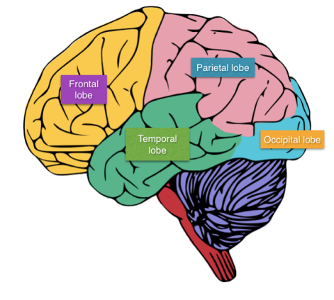
The cerebral cortex can be divided into four lobes. These are as follows:
Frontal lobe
Parietal lobe
Occipital lobe
Temporal lobe
You can remember the names for this using the mnemonic:
Freud Tore his Pants Off.
(I recommend you check out the blog post on brain directions before reading this next part).
Image: adapted from clker.com
So, using what we know of brain directions, we can use that terminology to say how the different lobes relate to each other in location:
For example, the frontal lobe is dorsal to the temporal lobe and rostral to the parietal lobe.
You can use something called the fist mnemonic to help you remember where all the lobes sit in the brain.
First, form a fist with your hand.
Then we’ll say that your thumb/thumb knuckle represents the temporal lobe.
Next, we’ll say that from the knuckles at the bottom of your fingers down to the front of your fist will form the frontal lobe.
Then from those finger knuckles down to about half way down the back of your hand represents the parietal lobe.
Then from half way down the back of your hand to the start of your wrist forms the occipital lobe.
We can also now use what we’ve learnt about sulci and gyri to know that the lateral fissure/ sylvian fissure divides the frontal lobe from the temporal lobe. Also, the central sulcus divides the frontal and parietal lobes.
Major lobe functions
Okay, now let’s take a quick look the major functions of each lobe within the cerebral cortex:
(Major apologies for the quality of these images. One day, I will remake these in a higher resolution!)
Extra information on the frontal lobe
Since the frontal lobe thinks it’s the captain of the show, naturally, it would want us to talk in-depth about all the individual components that make it up. So, making up the frontal lobe, we have the:
Prefrontal cortex
Premotor cortex
So, the prefrontal cortex’s jobs include:
Strategy
Making judgements
Planning
The prefrontal cortex is the true captain here. For example, Apple, as a company, may make decisions on what new product it creates. However, it’s the Chief Executive within the company who will actually make the final decision on what goes into the final product.
In this instance, the frontal lobe (company) makes the decisions. However, it is the prefrontal cortex (Chief Exec.) that makes the decisions and produces strategies and does all the planning.
The premotor cortex has the job of being in charge of something called the primary motor cortex, which you can read all about down below.
Primary areas of the cerebral cortex
There are 4 primary areas of the cerebral cortex:
primary somatosensory cortex (touch)
primary visual cortex (sight)
primary auditory cortex (hearing)
primary motor cortex (movement)
The…
primary somatosensory cortex
primary visual cortex
primary auditory cortex
…RECEIVE information from the SENSES.
The…
primary motor cortex
…SENDS information to the MUSCLES in your body.
Kind of like how a motor in a car makes the car move, your primary motor cortex tells your muscles to get moving.
Also, all the senses and motor signals are contralateral (e.g. the right eye’s connected to the…left hemisphere, the left eye’s connected to the…right hemisphere). The only exception to this is the sense of taste and the sense of olfaction (fancy word for smell).
The sensory association areas of the cerebral cortex
There are 3 sensory association areas:
Somatosensory association cortex
Auditory association cortex
Visual association cortex
The primary areas of the cerebral cortex (discussed above) send information to these 3 sensory association areas of the cerebral cortex for them to analyse.
So, it goes a little something like this…
For reference,
The somatosensory association cortex is located in the parietal lobe.
The auditory association cortex is found in the temporal lobe.
The visual association cortex covers both the temporal lobe AND the occipital lobe (because it’s greedy).
You can remember that the visual association area covers move than one lobe by remembering that when someone is being greedy and begging for lots and lots of food, they are said to have “eyes bigger than their belly”. Now if you extend this and remember a greedy person with huge eyes, you should remember that the visual association cortex is a greedy so-and-so that gobbles up, not one, but two lobes of the cerebral cortex.
The Basal Ganglia
The basal ganglia is essentially just a gang(-lia) of cell bodies (nuclei) living it up in your telencephalon.
The hoodlums that make up this gang-lia include:
caudate nucleus
putamen
globus pallidus
Now, the caudate nucleus and the putamen really come as a pair. Collectively, they are known as the striatum.
The globus pallidus is like the mafia boss of this gang-lia, with the striatum duo as his two lackeys. This is because the striatum receives all of the information (like lackeys working on a job for their boss) then sends this to the globus pallidus.
Image: stickmen images from canva.com
The functions of the basal ganglia include:
being in charge of movement control (intentional movement – NOT reactions)
being in charge of reward systems
motor learning
Although we said the frontal lobe of the cerebral cortex makes decisions on where we move to, etc – that is true – but what really happens is that the frontal lobe tells the basal ganglia (+ other parts of brain) where it reckons we should go and the basal ganglia then works (with other brain parts) to ensure that that movement is carried out effectively.
Motor learning is basically when your brain learns how to perform a series of actions so well that you don’t even need to think about what you’re doing to perform those actions anymore. An example would be when you enter your password on your phone. After a while your brain will have remembered the pattern that your thumb needs to move in without you even having to think about it. Another example would be when you drive a car. After a while of driving, your brain will learn in what order it needs to press the pedals and move the gear stick in order to get the car moving without it even crossing your mind.
If you want a way to remember the basal ganglia’s jobs, imagine it as a mafia gang that smuggles illegal drugs. They control the intentional movement of certain cargo (drugs) and being part of their gang could give certain financial rewards. Also, you can imagine that members of this mafia gang must be good getaway drivers, so anyone looking to join the gang must learn how to drive a motor vehicle fairly fast.
Image: adapted from cliparthut.com
If you have a lesion (big cut) in your basal ganglia, it can lead to the following disorders:
Parkinson’s disease
Huntington’s disease
(Side note: please don’t set up your own mafia-style gang…even if a job as a drug lord may pay better than whatever shelf-stacking job we all manage to find at Tesco after we leave university. R.I.P.)
Okay, and finally we move on to the last component of the telencephalon, the limbic system.
The Limbic System
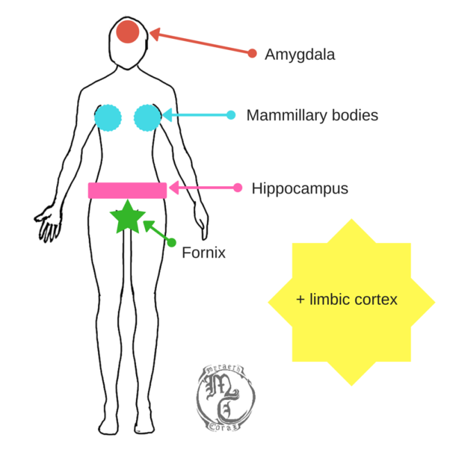
This is made up of the:
limbic cortex
amygdala
hippocampus
fornix
mammillary bodies
The general job of the limbic system includes: learning + memory
The Amygdala
The amygdala’s unique job is to help us learn what to fear by encoding certain stimuli as scary.
You could imagine Fear, a character who embodies the emotion of fear in the movie “Inside Out”. When Fear senses that the main character, Riley, has encountered something scary, he identifies this thing as scary and then later reminds her that this thing is scary the next time that she encounters it.
You could imagine that Inside Out’s Fear has a sister called Amy G. Dala who acts exactly as he does.
The Limbic Cortex
The limbic cortex looks a little bit like a sting ray when looked at from below, so it may help to remember the limbic cortex as the bit that looks like a sting ray.
“Limbus” actually means border. This is because the limbic cortex provides a border around the midbrain (as seen in top picture).
The limbic cortex also contains something called the cingulate gyrus (“sing yew lett“). The only tip that I have for remembering this is to perhaps imagine a young, single sting ray down at his local nightclub gyrating his hips in an odd dance in an attempt to attract a mate.
The limbic cortex has an important role in making sure that you avoid negative stimuli. So, you could imagine a string ray who tends to avoid the bigger, stronger males in the nightclub, who might want to pick a fight with him for accidentally hitting on their girlfriend.
Image credit: Clker.com
The Hippocampus
It’s name translates to “sea horse” because this little guy supposedly looks like a little sea horse in your brain.
This little sea horse sits in your temporal lobe.
His jobs include:
moving those short-term memories into the long-term memory (consolidation)
spatial navigation (so you remember the way back home after you leave the house…no bread crumbs needed here)
context
To remember this, imagine a sea horse who is also a taxi driver. Taxi drivers have to know MILLIONS of routes throughout the area in which they operate. Therefore, they must have good spatial navigation so that they can remember how to get to various places.They must also pay good attention to their current context, i.e. what’s going on around them, so that they can be safe when out on the road at rush hour.
Image: seahorse image adapted from cliparthut.com. Car image adapted from canva.com
How to remember the components of the limbic system
The limbic cortex is easy to remember – it’s the limbic system so limbic cortex should be straightforward.
As for the rest of the limbic system, try to imagine a generic image of a woman. This generic woman will be used to represent the limbic system.
Stereotypically, women are thought to be more empathetic than men. We’ve already discussed that the amygdala is very important for emotion in the brain. In fact without it, you’d feel a lack of empathy.
Therefore, when we think of the generic woman’s emotive brain, we can link it to the amygdala.
The term “mammillary bodies” literally means “breast shaped”. Therefore, the woman’s breasts can be representative of the mammillary bodies in the limbic system.
The hippocampus can be represented by the woman’s hips (HIP-pocampus).
The fornix sounds like the cervix. The cervix is the name given to the lower part of the uterus in a female’s reproductive system. Thus, the generic woman’s cervix can represent the fornix.
So you should be trying to remember this:
Image credit: adapted from MyraethCorax @ DeviantArt.com
Forebrain: Diencephalon’s structures
Before we move on, let’s remind ourselves of our brain divisions:
We now know about all the structures in the first subdivision of the forebrain, the telencephalon. Now let’s look at the structures within the second subdivision of the forebrain, the diencephalon.
First things first, you can remember that the diencephalon is the second division of the forebrain because the prefix “di” means “two“, therefore, the diencephalon must come second.
Within the diencephalon, we have the:
Thalamus
Hypothalamus
Thalamus
The thalamus is formed of one lobe in each hemisphere – two lobes in total. These two lobes are separated by the massa intermedia.
The thalamus is essentially the brain’s post office. It receives sensory information and sends this to the cortex in a relay. Each nuclei within the thalamus deals with a certain type of sensory information.
The nuclei of the thalamus are:
lateral geniculate nucleus
medial geniculate nucleus
ventrolateral nucleus
Image: eye, shoe and sound icon from canva.com
And the same info as a table:
So:
the retina projects visual info to the lateral geniculate nucleus in the thalamus, which then sends it to the primary visual cortex in a relay.
the inferior colliculus projects auditory info to the medial geniculate nucleus in the thalamus, which then sends it to the primary auditory cortex in a relay.
the globus pallidus, cerebellum and substantia nigra project motor info to the ventrolateral nucleus in the thalamus, which then sends it to the primary motor cortex in a relay.
(If words like “inferior colliculus” are freaking you out – chill! All will be explained soon – just keep reading).
Hypothalamus
When thinking about the thalamus and the hypothalamus, I like to think of them as a pair of brothers. The thalamus is the responsible one who has a job as a postman, delivering information to the cortex. Whereas, the hypothalamus is the thalamus’ hormonal teenage brother.
Hypothalamus’ functions:
controls the autonomic nervous system
controls the endocrine system
These 2 functions mean two things for the hypothalamus. First of all, let’s start with it’s job of controlling the endocrine system.
The endocrine system is the system that controls the release of hormones throughout the body. The hypothalamus does this by having a link to the pituitary gland. The hypothalamus basically tells the pituitary gland which hormones to release.
Think back to your teenage days, specifically, every time your hormones sent you into an irritated, sulky mess. Well, this little guy is the culprit.
Also, every time you get stressed and your heart begins to race – that’s this little guy’s fault too.
This brings us to his second job, controlling the autonomic nervous system. The autonomic nervous system is the system that kicks in when you’re in a fight or flight scenario, i.e. your heart races, your pupils dilate, your digestion shuts down, etc.
Your autonomic nervous system is switched on via the release of certain hormones.
This means that if the hypothalamus gets all stressed about breaking a nail, it would then tell the pituitary gland to release certain hormones into your bloodstream and next thing you know your heart is racing, adrenaline is surging through your body and you’re feeling like you’re about to fight a bear.
In summary, it’s good to think of the hypothalamus as a moody, hormonal teenager who overreacts to a lot of things (although, all the hypothalamus is trying to do is help you survive). Also, the hypothalamus means “under” which helps the little brother image.
You could also think of the hypothalamus as a primitive ape who reacts in a fight or flight way quickly before rational thinking has change to kick in.
Midbrain: structures
Again, let’s have a recap:
Now the midbrain differs from the forebrain and hindbrain as it has no subdivisions. Just remember that the one in the middle doesn’t have subdivisions.
The midbrain or mesencephalon has the following structures:
Superior colliculi (singular: superior colliculus)
Functions:
To remember the separate functions of each, think of how we as humans have evolved in a way that means that our sense of sight is far more precise than our sense of hearing. We can use our sight to pinpoint the location of a predator, even in low level light. Whereas, our sense of hearing could not accurately tell you the location of a predator.
In this instance it could be argued that the sense of sight is superior to the sense of hearing.
Tegmentum
This is made up of the:
reticular formation
periaqueductal grey matter
red nucleus
substantia nigra
Functions:
Bonus-
The red nucleus is actually called red due to its high iron content.
Damage to the substantia nigra can result in the development of Parkinson’s disease
Hindbrain: Metencephalon’s structures
Finally, we move onto the hindbrain or the rhombencephalon. The first subdivision of the hindbrain is the metencephalon.
Let’s once again, recap:
The metencephalon’s structures include:
Cerebellum
Pons
Cerebellum (“sair a bell um”)
The cerebellum is the ball-like lump that sticks out at the back of your brain.
Like your cerebrum (main part of your brain), the cerebellum has a left and a right hemisphere.
The cerebellum is also more densely packed than the cerebral cortex, which means it has more folds (+looks more wrinkly).
Cerebellum’s jobs:
Coordination of movement
However, recent studies have found that the cerebellum may also perform these jobs too:
timing
rhythm
cognition
attention
Pons
Pons’ jobs:
control of sleep and arousal state
carrying info from the cortex to the cerebellum
Pons literally means “bridge” and you can use this to remember how the pons acts as a bridge to allow the information from the cortex to reach the cerebellum.
The name pons is similar to the name of the greek god, Pan, who was the god of sexuality (amongst other things).
You could use this to answer the question: What is one of the functions of the pons?
Just think pons> Pan > god of sexuality > arousal > arousal state > controlling of arousal state + sleep.
Or perhaps you could imagine the image of a generic greek god and say that this is a god called Pons.
This new greek god is the master of sleep and wakefulness. (Hypnos is actually the greek god of sleep BUT for the purpose of remembering this, we can pretend his name is Pons).
So when you’re asked a question about the pons, think: ah, yes, Pons – the greek god of sleep and wakefulness who controls when people sleep and wake up. Boom.
Hindbrain: Myelencephalon’s structures
Finally, we arrive at the second division of the hindbrain/rhombencephalon, the myelencephalon.
Let’s have a final recap:
There’s only one structure within the myelencephalon (and it’s my personal favourite):
medulla oblongata
Medulla oblongata
Jobs of the medulla oblongata:
regulating the cardiovascular system
regulating respiration
regulating skeletal muscle tonus
vital reflexes
Going down the list:
Regulating the cardiovascular system
The cardiovascular system is the system that keeps your heart pumping. This is very important.
Regulating respiration
Image credit: Clker.com
Apologies for echoing the voice of your GCSE Biology teacher but RESPIRATION DOES NOT MEAN BREATHING. Respiration means converting glucose into energy. Energy that you need to go about your daily living. This is also very important.
Regulating skeletal muscle tonus
Image credit: Clker.com
Skeletal muscle tonus basically refer to ability of the muscles to hold your body/limbs in certain positions. This is also very important.
Vital reflexes
Image credit: Clker.com
Every time you: cough, sneeze or vomit, you’ve got this little guy to thank.
Don’t be so ungrateful, this little guy is saving your life every time this happens, as by doing this he stops viruses and other nasties getting into your body. Thanks, Medulla.
To remember the functions of the medulla oblongata we’re going to do a little dance.
Put your hands together over your heart and raise them on and off your chest repeatedly like a heart beating. This represents the regulation of the cardiovascular system.
Now run on the spot. This represents the regulation of respiration (so you have the energy to keep running).
Now stop and flex your muscles. This represents the regulation of skeletal muscle tonus.
Finally, do a big sneeze! This represents the vital reflexes.
Do this on repeat whenever you have a spare moment (you can run through the actions in your head rather than acting them out) and eventually, it’ll stick in your head.
Other medulla info to know:
The medulla oblongata contains gaps. These gaps allow substances to pass through. As mentioned a moment ago, if a toxic substance enters the medulla, good ol’medulla sends out a signal that makes you vomit/sneeze/cough in order to get rid of the nasty toxic substance. What a good guy.
So next time you complain about having a sneezing fit, just remember that that’s your medulla trying to save your life.
Damage to your good guy medulla can result in something called locked-in syndrome. This syndrome includes patients not being able to move their body, apart from their eye muscles.
So look after your good guy medulla. He’s constantly trying to protect your life, after all.
Phew! You’ve made it to the end of this extra-long post! Here’s a present for your efforts. It’s a hierarchy chart of all the divisions, subdivisions and structures that we just went through.
The nervous system is divided into two main structures:
Central Nervous System (CNS)
Peripheral Nervous System (PNS)
The Central Nervous System (CNS)
This contains the:
Brain
The spinal cord
The Peripheral Nervous System (PNS)
This structure contains the nerves.
These nerves form two types of pathways:
Motor pathways (these go to the muscles)
Sensory pathways (these go to the brain)
Image credit: Taken from Human Body Colouring Book (2015), Amber Books Ltd.
The Brain: basics
The brain is physically made up of:
Cerebrum (“sair ree brum”)
Cerebellum (“sair a bell um”)
Brain stem
The cerebrum is the main structure of the brain.
The cerebellum is the ball that sticks out at the back of the brain.
The brain stem is the long stick at the bottom of the brain that hooks the brain up to the spinal cord.
Brain basics: hemispheres
The brain has two hemispheres, one left hemisphere and one right hemisphere.
Image credit: Taken from Human Body Colouring Book (2015), Amber Books Ltd.
A lot of brain function is what we call contralateral function.
For example, the left hemisphere controls movement on the right side of the body and vice versa. One hemisphere controls the opposite side of the body. Like when someone contradicts someone else, they take the opposite viewpoint.
If the opposite were true, this would be called ipsilateralfunction.
This is when one hemisphere controls movement on the same side of the body. For example, if the left hemisphere controlled the left side of the body.
The Corpus Callosum (“Core pus ka low sum”)
This is the bundle of fibres that keeps the two hemispheres of the brain connected. It’s name translates to “hard body”.
Any pathway that connects a left and right hemisphere together is known as a commissure.
The corpus callosum is an example of a commissure and is the largest one in your body.
Grey and white matter
Image credit: Clker.com
The brain contains both grey matter and white matter.
Grey matter refers to cell bodies and dendrites that make up the main structures within the brain.
Whereas, white matter refers to myelinated axons that help transmit information around different part of the brain.
Chopping the brain up
Not saying that you would, but if you were to chop your annoying little brother’s brain in half then you’d have many options on exactly how to cut the brain.
Frontal (transverse/coronal)
Once you’ve somehow extracted his brain, you could give it a frontal cut.
This is parallel to the forehead.
It’s called a frontal cut because you get to see the brain from the front.
Just to be annoying, some people also refer to it as a coronal cut. You can remember this by thinking about a blade hitting the crown of your head and going straight through (ow!). A coronal cut would be left behind and coronal sounds a little like crown.
Even more annoyingly, some people also refer to it as a transverse cut. (I wish they’d make their mind up!). You can remember that it’s called a transverse cut because transverse means “to extend across something” and a transverse cut extends across the front of the brain.
Sagittal cut
To perform this cut, you’d send the knife right between your annoying brother’s eyes and cut his brain straight down the middle. When you’d finished, you’d be able to see one side of his brain (please don’t try this at home).
Image credit: Taken from Human Body Colouring Book (2015), Amber Books Ltd.
A way to remember this is to think of the star sign Sagittarius. This star sign takes the form of an archer and when archers stand to shoot an arrow, they turn their head to the side. If it helps, sagittal actually translates to “arrow”.
Image: from computerclipart.com
Horizontal cut
This is a cut that is parallel to the ground. Imagine you’ve got your annoying brother pinned to the floor, lying on his stomach with his chin on the carpet. To avoid making a mess and ruining your mum’s cream carpet, you’ll want to make one neat cut through the brain. The best way to do this is to keep your blade paralell to the floor, like you’re cutting the top off of pumpkin.
Image credit: Human Body Colouring Book (2015), Amber Books Ltd.
If violent analogies aren’t your thing, then you could also think about the horizon; a theoretical line that sits parallel the Earth’s surface, separating the ground from the sky.
DISCLAIMER: This blog is not responsible for any death or injury. This blog does not condone any violent behaviour. Please do not actually murder your brother/anyone.
Protection of the nervous system
The brain uses the following to defend itself:
ray guns
death beams
nukes
Okay, that may not be true (but how cool would that be?!).
Actually, the brain uses a structure called the Meninges (“men in jees”) to protect itself.
The meninges are formed of 3 layers of tissue. These 3 layers protect the brain and the spinal cord (so, all of the CNS).
The 3 layers of the meninges are called:
Dura Mater – (Remember it because, it must be Durable to be the top layer of the meninges)
Arachnoid Membrane – (Remember it because, it’s the spider membrane that webs the other two layers together, since arachnoid sounds like arachnid)
Pia Mater – (Remember it because, since it’s the bottom layer of the meninges, you’d really have to Pia in to get a good look at it!)
Image source: Antranik.org
Inflammation of the meninges is known as meningitis, which is a deadly illness that you may have heard of.
The meninges float in something called cerebrospinal fluid (CSF). CSF is a clear liquid that fills the subarachnoid space (the space between your brain and the meninges; see picture).
CSF’s functions include:
shock absorption (in case of head injury)
buoyancy
reducing the weight of the brain (so you can actually hold your head up!)
The Ventricular System
Your brain contains four interconnected structures called ventricles (‘ven trik uls‘) , which translates to ‘little bellies‘.
These ventricles are all filled with cerebrospinal fluid (CSF).
The ventricular system includes two lateral ventricles, with one in each hemisphere. Lateral means on the side of an object. So, you can remember that the brain has one lateral ventricle on each side.
This system also contains: a third ventricle, a fourth ventricle and a cerebral aqueduct.
Aqueducts were large, bridge-like structures used by the Ancient Romans to transport water. Therefore, you could remember this by thinking of the third and fourth ventricles as two little cities. The cerebral aqueduct then acts as a mechanism for transporting the CSF from the third ventricle to the fourth ventricle, like transporting water between two cities.
These can all be seen here:
This picture shows the production and movement of CSF within the ventricular system:
Here’s the same information again in a list format for those who prefer lists:
Here’s the process again with some ways to remember the steps:
A membrane known as the choroid plexus filters the blood to produce CSF.
Remember this because: the choroid plexus sounds like your solar plexus which is located in the pit of your stomach/gut. Your gut also does a lot of filtering when it filters out the waste products from the food that you eat.
Therefore, just think – choroid plexus – sounds like solar plexus – gut – the gut filters stuff – the choroid plexus filters blood into CSF!
This CSF is then stored in the lateral ventricles.
Remember this because: you can picture the lateral ventricles like a pair of testicles – there’s a left one and a right one. The difference being that, where the testicles store sperm cells until it’s needed, the lateral ventricles store CSF until it’s needed.
The CSF then travels down into the third ventricle.
Then through the cerebral aqueduct.
Then through the fourth ventricle and finally travels down the spinal cord.
Remember this because: sticking with the testicles analogy (because that’s certainly going to stick in your mind), the third ventricle, cerebral aqueduct and fourth ventricle can be thought of as being like the shaft of a penis (stay with me here).
In a penis, when the sperm cells are needed they travel from the testicles, where they are being stored, down through the shaft.
Similarly, in the ventricular system, when the CSF is needed, it travels from the lateral ventricles, where it is being stored, down through the third ventricle, cerebral aqueduct and fourth ventricle.
As an alternative, you could imagine that the lateral ventricles are like little factories that work collaboratively to create the CSF. The CSF is then stored in the warehouses of the factories until it’s needed. When the CSF is needed, the CSF is transported from the warehouses (perhaps in an Amazon Prime van) to city A (the third ventricle) and city B (the fourth ventricle). However, to get to city B, the little van has to travel across a bridge (the cerebral aqueduct). When the CSF has been delivered to city B, the little van then travels down the spinal cord to complete the rest of his deliveries.
(Hannah from 2020 here. I originally wrote this blog post 3-4 years ago when I was an undergraduate. Upon rereading this, I admit, the penis analogy is a bit weird. However, I’ve decided to leave it in as I think that it succeeds in creating a memorable analogy, which was my goal here. I have also added an alternative analogy, just in case penis analogies aren’t your cup of tea).
Why does this happen though?
The purpose of the ventricular system is to carry waste away from the brain (and down the spinal cord into the rest of your body…so, it’s like the brain is pooping on you…ew!).
The Blood-Brain Barrier
These are semipermeable barriers. Permeable means “lets stuff through” – so semipermeable means that they only let SOME stuff through.
Blood-brain barriers allow lipid soluble substances (fat soluble, substances that dissolve in fat) to pass through as they wish.
However, substances with LARGE MOLECULES aren’t allowed to pass through easily and must be actively transported through the walls instead.
Imagine the blood-brain barriers are all nightclub owners. They are also all jerks. They only let small skinny substances (small lipid soluble substances) to pass through the doors into their club.
They don’t let larger substances (with larger molecules) to pass freely through them and into the club.
Instead, any substances with larger molecules must be actively transported past the barrier. In the jerky club owner analogy, you can think of this as that the larger substances must offer the jerk blood-brain barrier a bribe, in order to get past and be “actively transported” into the club.
Image source: molecule image from Clker.com
They are similar to capillaries in the rest of your body however, unlike those, blood-brain barriers do not contain gaps.
DISCLAIMER: I do not condone any form of body-shaming. Be nice to each other. Do not be a jerk blood-brain barrier.
But why are the blood-brain barriers such jerks?
Well they keep out larger substances to:
Maintain a stable environment
To give protection from dangerous chemicals
(like a bouncer does when he keeps violent people out of the club to maintain a somewhat nice environment inside)
Think you’ve got it?
Test yourself here.
(Image credit for image of frontal brain cut: “Human brain frontal (coronal) section: #24 is lateral sulcus” by John A Beal, PhD Dep’t. of Cellular Biology & Anatomy, Louisiana State University Health Sciences Centre Shreveport. No copyright intended. Used in accordance with the CC-BY 2.5 license. Disclaimer: I do not own this image.)


_section_description_2.JPG)


